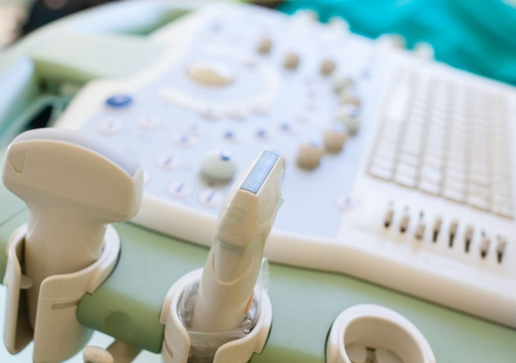
In the realm of veterinary medicine, sound wave imaging, or ultrasound, has emerged as a cornerstone technology, offering unparalleled insights into the health of pets. As a trusted partner in pet ultrasound, sound wave imaging provides veterinarians with a non-invasive, high-resolution tool for diagnosing and monitoring a wide range of conditions. This article explores the significance of sound wave imaging in veterinary care and its role as a reliable partner in pet health.
Understanding Sound Wave Imaging
Sound wave imaging utilizes high-frequency sound waves to create real-time images of internal structures. A transducer emits sound waves that penetrate the body and reflect off various tissues and organs. The echoes are then captured and transformed into visual images by a computer. This imaging technique is prized for its safety, as it does not involve ionizing radiation, making it ideal for both routine and complex diagnostics in animals.
The Benefits of Sound Wave Imaging for Pets
- Non-Invasive Examination: One of the primary advantages of ultrasound is its non-invasive nature. Unlike surgical procedures, ultrasound allows for internal examination without the need for incisions. This minimizes stress and discomfort for pets, facilitating more frequent and comprehensive evaluations.
- Real-Time Imaging: Ultrasound provides real-time imaging, allowing veterinarians to observe internal organs and structures as they function. This dynamic view is crucial for assessing organ movement, blood flow, and other physiological processes, leading to more accurate diagnoses and treatment plans.
- High-Resolution Details: Modern ultrasound technology offers high-resolution images that reveal detailed anatomical structures. This precision is invaluable for diagnosing conditions such as tumors, organ abnormalities, and structural defects. High-resolution imaging enhances the ability to identify and monitor changes in soft tissues and organs.
- Safety and Versatility: Ultrasound is a safe imaging modality, with no exposure to radiation. It can be used across a wide range of clinical scenarios, from routine wellness exams to emergency assessments. Its versatility makes it suitable for evaluating various conditions in different organs, including the abdomen, thorax, and heart.
Applications of Sound Wave Imaging in Veterinary Care
- Abdominal Ultrasound: Abdominal ultrasound is commonly used to assess the liver, spleen, kidneys, bladder, and gastrointestinal tract. It aids in diagnosing conditions such as liver disease, kidney stones, tumors, and gastrointestinal obstructions. By providing detailed images of these organs, ultrasound helps veterinarians develop targeted treatment plans.
- Cardiac Ultrasound (Echocardiography): Echocardiography is a specialized type of ultrasound used to evaluate heart function and structure. It is essential for diagnosing heart diseases, such as congestive heart failure, valvular disorders, and congenital heart defects. Echocardiography provides detailed information about the heart’s chambers, valves, and blood flow.
- Thoracic Ultrasound: Thoracic ultrasound allows veterinarians to examine the lungs, pleura (lung lining), and other structures within the chest cavity. It is useful for detecting conditions like pleural effusion (fluid accumulation), lung tumors, and abnormalities in the chest cavity.
- Pregnancy Monitoring: Ultrasound is a key tool for monitoring pregnancies in female pets. It helps confirm pregnancy, assess fetal development, and detect potential complications. Regular ultrasound evaluations ensure the health of both the mother and her developing puppies or kittens.
- Guided Procedures: Ultrasound can guide various procedures, such as biopsies and fluid aspirations. By visualizing internal structures in real-time, veterinarians can perform these procedures with greater accuracy and reduce the risk of complications.
The Role of Veterinary Professionals
Veterinary radiologists and ultrasound technicians play a crucial role in the effectiveness of sound wave imaging. They are responsible for operating the ultrasound equipment, interpreting the images, and providing detailed reports to referring veterinarians. Their expertise ensures that the imaging process is conducted accurately and that the results are used to guide effective treatment decisions.
Advancements and Innovations
The field of sound wave imaging continues to evolve, with advancements in technology enhancing its capabilities. Innovations such as 3D and 4D ultrasound provide even more detailed views of internal structures, while contrast-enhanced ultrasound improves visualization of blood vessels and tissues. These advancements contribute to more precise diagnostics and better outcomes for pets.
Conclusion
Sound wave imaging stands as a trusted partner in pet ultrasound, offering a non-invasive, high-resolution tool for comprehensive veterinary care. Its benefits, including real-time imaging, detailed anatomical views, and safety, make it an indispensable part of modern veterinary practice. From routine examinations to specialized diagnostics and guided procedures, ultrasound plays a vital role in ensuring the health and well-being of pets. As technology advances, sound wave imaging will continue to be a cornerstone of veterinary diagnostics, helping to provide the best possible care for our beloved animal companions.


